Measurement of bone mass density Bone Mineral Density-BMD is a measurement of bone density values based on different parts, helping to determine what level of bone health is strong and whether there is osteoporosis.
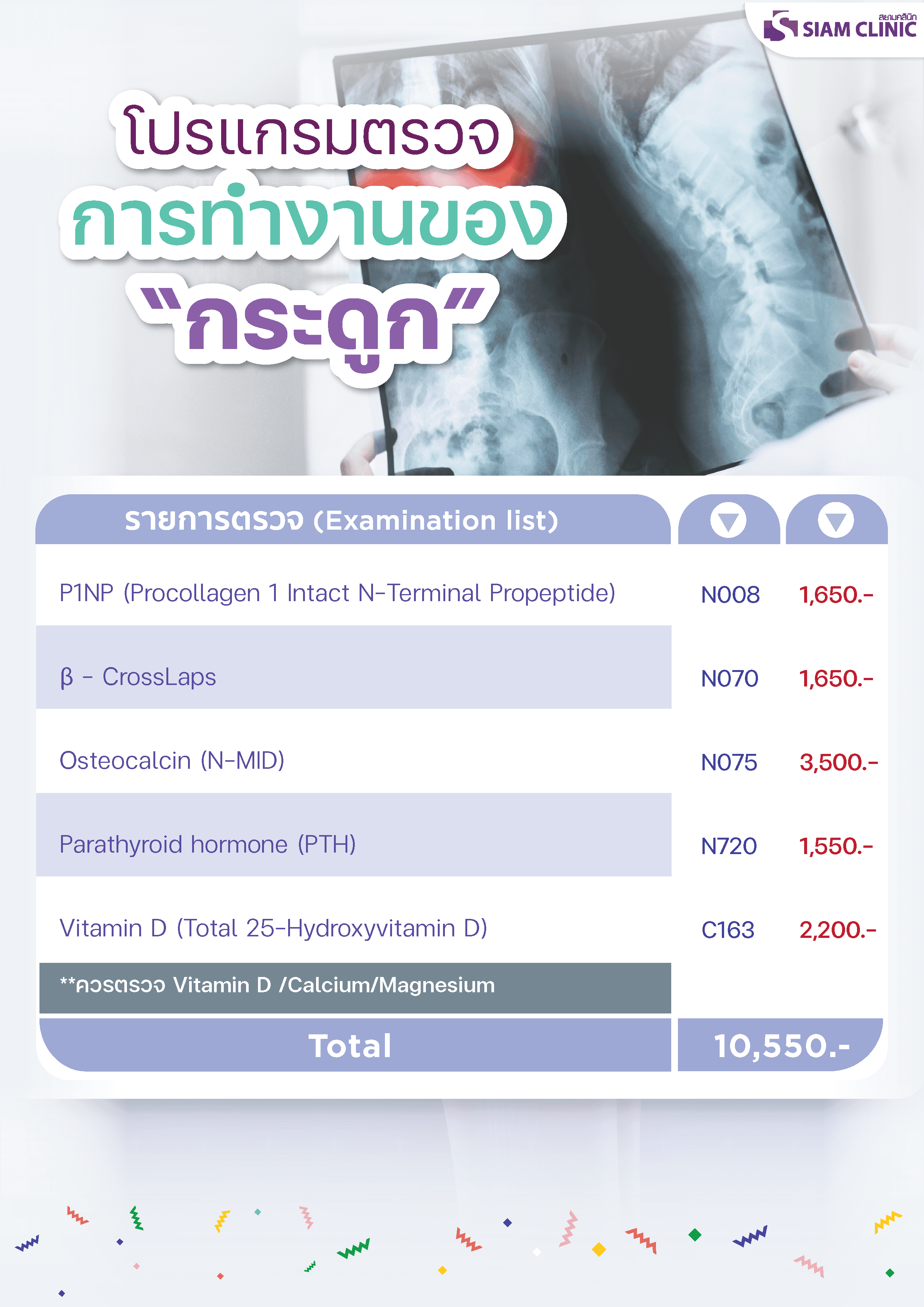
List of bone function examinations
- P1NP (Procollagen 1 Intact N-Terminal Propeptide)
- β – CrossLaps
- Osteocalcin (N-MID)
- Parathyroid hormone (PTH)
- Vitamin D (Total 25-Hydroxyvitamin D)
**Vitamin D /Calcium/Magnesium should be examined.
Is osteoporosis really scary?
Osteoporosis refers to the fact that the patient’s bones become less dense, causing them to break easily. Diagnosis of the disease uses information from history, detailed physical examinations. Blood tests and radiographs for diagnosis and differential analysis
Specifically, differential diagnosis of vitamin D deficiency. Calcium deficiency disease, cancer Parathyroid gland disease and other co-occurring diseases, physical examinations and general blood tests showed no abnormalities. Radiographs showed bones thinner than usual, particularly at the ends of the limbs. Sciatica and spine Thinner bones are common. No bone damage was observed. The fracture site has an increased radiation opacity due to bone stacking.
Bone density tests, as described above, are usually found to be lower than normal, based on a t score if it is lower than normal but still higher than -2.5. Blood tests to observe the development and function of bone cells that help diagnose and detect changes are available for treatment: Parathyroid hormone levels, dosage test Bone or bone decomposition and regeneration examination.
This osteoporosis is most commonly found in female patients of postmenopausal age. This is followed by patients who have been taking steroids for a long time. In male patients, it is found in people who regularly drink a lot of alcohol. Osteoporosis patients often develop fractures, followed by pain. Deformity and chronic pain Care guidelines should classify people at risk of developing osteoporosis and fractures. The results of the OSTA (Osteoporosis Self-Assessment Tool for Asians) study were calculated from the formula of 0.2 x (body weight). Reed.– Age, year) is an index that shows the risk of osteoporosis. If the value is obtained less than – 4, the patient is at high risk. If it is between – 4 and – 1, the patient is at moderate risk. 1 Low risk patients
Osteoporosis, so what happened?
In general, patients with osteoporosis in the early stages do not have pain, although the bones are much thinner. But when these patients are born, fractures are formed. It has been found that patients often experience chronic pain, especially fractured or collapsed spines. Patients often have a lot of back pain and chronic pain that cannot be explained by the nature of the entire injury.
Some groups of patients have numbness. Weakness and a feeling of touch that is wrong, followed by chronic pain and depression. This chronic pain disorder in patients with osteoporosis and fractures is of interest to scientists and doctors. It brings about a study to determine the cause to lead to better treatment.
The cause of pain in patients with osteoporosis and fractures is due to:
1) Pain from injuries, fractures. Fractures injure the various tissues surrounding the bone. Death of the end of the bone occurs. Bone membranes and often experience muscle injuries surrounding the bone. These stimulate the sensory nerve endings, then carry the signals of studs to the spinal cord to the brain, causing pain.
2) Pain from inflammation Fractures cause blood clots to accumulate around the fractured bone. The blood clot stimulates the surrounding tissues, causing inflammation, and in cases where the fractures through the joints cause inflammation in the neighboring joints. In patients who cannot provide treatment by pulling the bones into place, the joint may deteriorate faster than it should. It has the effect of causing chronic pain.
3) Pain from disturbed or injured nerves, broken bones, especially broken vertebrae may press or disturb the spinal cord. Nerves or nerve roots In the early stages, it is pain from nerve injuries. If pressing or disturbing nerves cannot be resolved, it can lead to chronic neurodegenerative syndrome or pain syndrome. Patients have pain in combination with abnormal functioning of the nervous system, such as osteoporosis and fractures in the extremities of the wrist bone, which often cause chronic pain syndrome and knuckle sticks. And patients with spinal fractures from osteoporosis may experience pain to the chest. The abdomen and legs together with various sensory disorders, as found in chronic pain syndromes of the nervous system.
In osteoporosis patients, abnormalities in the coordination between the nerves that feed the bones and the bone cells are observed.
In normal conditions. The nervous system regulates the functioning of various cells of bones. On the other hand, the various cells of the bone feed the nervous system through the nerves that feed into the bones. The shape of bone cells is like a nerve cell and works, receiving commands from the nervous system through nerves that come into direct contact with bone cells. Subsequently, osteocyte bone cells activate to bone-breaking cells. Remove any injured or unnecessary bones and instruct the cell to rebuild the bone. There is evidence to believe that bone cells are cells that receive information from the bones and surrounding solutions and then pass them on to the nerves in another place, creating a balance.
In the elderly, the bone cells are aging down. Nerve cells increase their functioning more, which can be the beginning of excessive functioning and the walls of nerve cells become more sensitive. When a fracture occurs, there may be an abnormally severe nervous cell response, which is the source of chronic neurological pain.
4) Muscle pain caused by deformity of broken bones. This causes the surrounding muscles and ligaments to work harder in order to achieve the best balance possible. It causes pain and can cause muscle tone.
Degeneration occurs early.
How is osteoporosis treated?
Viewing and treating osteoporosis patients is aimed at preventing fractures. Key treatment guidelines include:
1) Osteoporosis education and correct treatment principles
2) Recommending practices to reduce the risk of fractures, both in the personal and home environments.
3) Proper administration of the body to maintain bone strength. This allows strong muscles to better support the bone system, as well as training the joint functioning of the musculoskeletal system and nervous system to help prevent falls, or if a fall, the patient may have better reflexes, help prevent severe falls and prevent fractures.
4) Use of walking aids and fasteners, bending and supporting the patient’s body and joint area to increase stability.
5) Eating symptoms with a disproportionately high nutritional value. Foods high in calcium are recommended and calcium should be given at a dose that produces calcium absorption of approximately 1000 mg per day, taken with a diet, and provide vitamins, especially vitamin D supplements, at a dose of 400 to 1200 units per day.
6) Medicinal treatment The use of the drug depends on the nature of the pain and pathology of the pain. The drugs given are divided into 3 groups:
6.1) Common pain relievers, paracetamol and anti-inflammatory drugs. The doctor considers choosing the appropriate medication for each inpatient. With this group of patients, they are usually of advanced age. There are several other complications. There is an easy chance of developing peptic ulcer when using anti-inflammatory drugs. In addition, patients often have to take several medications already.
6.2) Bone damage medications include:
6.2.1) Calcitonin
6.2.2) Bisphosphonates
6.2.3) Estrogen
6.2.4) Raloxifene
6.2.5) Parathyroid hormone
6.2.6) Vitamin K2
6.2.7) Vitamin K2
6.3) Medications relieve chronic pain from the nervous system.
Each drug has different indications, depending on the nature of the disease in each patient, with the principle of inhibiting bone destruction and encouraging the body to build a freckle in order to achieve stronger bones. It was determined by the supervising physician for a physical examination. Blood tests and bone density tests.
7) Surgical treatment The surgery aims to make the patient recover from the pain and return to their original life as quickly as possible. In patients with a fractured lower ulnar bone. It is preferable to pull the bones into place and damn them with splints, and then give the patients a speedy rehabilitation. In patients with fractures in the sciatica, treatment is preferred by surgically pulling the bone into place and damning it with metal. By choosing the right bone metal. It can hold thin bones well. In patients with a broken femoral neck, surgery is recommended to replace the prosthesis to reduce complications and the patient can return to his seat. Stand and walk quickly. In patients with spinal fractures and subsidence. If there is not much postponement or collapse, conservative treatment can be provided. By resting in the early stages, providing pain relief medications, providing regenerative medicine treatments in conjunction with the use of fasteners, bending and supporting the spine, but if the bone is very collapsed or the bone is broken, if it is preferable to inject bone substitutes or bone cement into the fracture site to connect the spine to a stable tremor. The patient can get up early. If a broken bone interferes with the spinal cord and or nerves, surgery may be performed to align the bone in place and metallurgically in combination with the bone transplantation in the fractured area, and then continue to provide medical and regenerative treatment.
How can osteoporosis be prevented?
Preventing osteoporosis must inevitably be better than treatment. People should receive sufficient amounts of calcium from childhood and young adulthood, avoid foods and substances that cause bone damage or cause the body to expel a lot of calcium from the body, such as drinking soft drinks. Drinking several glasses a day and drinking regularly should be exercise appropriate for your age and body shape. Proper exposure to sunlight. Treatment of chronic diseases such as diabetes to be close to normal. If there are problems, it is better to consult a doctor immediately.
Osteoporosis is a condition in which bone texture becomes thinner due to loss of bone mass. This happens gradually, especially in the spine, hip bones, and data bones. Generally, there are usually no signs until a fracture occurs, where the risk factors that cause osteoporosis include:
- aged
- female
- White and Asian ethnicity
- Have menopause before the age of 45.
- Thinning body structure and low body mass index.
- heredity
- Insufficient calcium intake.
- Lack of physical activity.
- Smoking, drinking alcohol, caffeine on a regular basis.
so The Osteoporosis Self-Assessment Tool for Asians (OSTA) is a osteoporosis risk assessment calculated based on age and body weight to detect the level of risk of osteoporosis and to treat and adjust behaviors to reduce future risks.
Examination of bone mass density. What is it?
Bone mass density examination It is a bone health check to see if it is at a normal level or if it is osteoporosis. The DEXA Scan is the most standard bone mass density measurement machine available today. Used in the diagnosis of osteoporosis.
Bone mass density
Bone mass density If the dosage is reduced, it is easy to risk fractures. The decrease in bone mass; It usually diminishes with increasing age. It can occur both in men and women at high risk after menopause, as well as other causes such as having a history of brittle bones breaking easily. Taking certain groups of medications for long periods of time, suffering from rheumatoid arthritis. Or overwork of the parathyroid glands, etc.
What to inform your doctor before receiving the service
- There is metal embedded in the body, such as bone iron insertion. Hip prosthetic joint Defibrillators, etc.
- Suspected pregnancy
- Have a history of treatment with mineral ingestion or opaque injections for diagnosis.
symptom
People with osteoporosis usually know once the symptoms have developed, but there are indications that can be observed in order to be treated early, and there are other indications that should be observed so that they can be treated in time.
- Wrist, arm, hip, and spine bones are easily fractured. Even mild bumps.
- Humpback or upper spine curved downwards
- Reduced height
- There may also be chronic back pain.
Bone mass density examination What are the advantages?
- This makes it known that the bones are still of normal density or have osteoporosis. How is it necessary to treat or strengthen bones?
- After the examination, you will know how to behave properly or what to take extra precautions.
Osteoporosis
Osteoporosis is a condition in which bone mass decreases. As a result, the bones lack strength, break easily. It is a disease that is more common in women than men, especially women in the first 5 years after menopause because of bone mass loss comparable to that of men older than men aged 5-10 years.
Who should be screened?
– Women over 45 years old or men over 60 years old
– Postmenopausal women
– Women who have had surgery on both ovaries before menopause
– People with a history of broken bones
– People with chronic pain in the back, waist, wrists
– People whose physical appearance of the outline has changed, such as a deflection back, shoulders cocked, or lowered.


